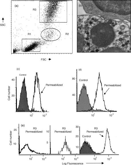
Figure 1.
Demonstration of cytoplasmic antigens using FACSLysing solution. (a) Dot-plot of forward (FSC) versus side angle light scatter (SSC) showing three main types of human peripheral blood leucocyte: R1 = lymphocytes, R2 = monocytes and R3 = neutrophils. (b) Transmission electron micrograph of leucocytes following fixation and permeabilization. Intact plasma and nuclear membranes of a lymphocyte (top cell) and a neutrophil (bottom cell) are visible. Cytoplasmic organelles are also intact. (c) Histogram analysis of neutrophils (R3) showing uptake of propidium iodide by control viable cells (solid area) compared with fixed and permeabilized cells (single line). (d) Histogram analysis of neutrophils (R3) showing binding of monoclonal anti-vimentin (intermediate filaments) by control viable cells (solid area) compared with fixed and permeabilized cells (single line). (e) Histogram analysis of lymphocytes (R1), monocytes (R2) and neutrophils (R3), showing binding of monoclonal anti-myeloperoxidase following fixation and permeabilization of cells (single line). This monoclonal antibody did not bind to viable cells (surface staining) as shown for R3 as a solid area.

