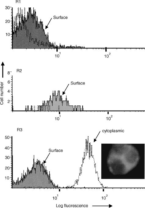
Figure 2.
Demonstration of cytoplasmic CD86 (B7.2) using FACSLysing solution. R1, histogram analysis of lymphocytes showing no significant binding of FITC-conjugated monoclonal anti-CD86 either before (solid area) or after (dotted line) permeabilization. R2, weak surface staining of monocytes for CD86 but no cytoplasmic staining observed. R3, strong staining of neutrophils for CD86 is evident (mean fluorescence intensity = 50), following fixation and permeabilization, as indicated by the dotted line. The overlay shows that surface staining (solid area) was not observed when viable cells were used in parallel. The photograph shows that when viewed under UV-light, fluorescence (FITC-anti-CD86) was observed predominantly within the cytoplasm of neutrophils with little or no nuclear staining observed. Similar results were obtained using monoclonal antibodies specific for CD20, CD21, CD22 and CD80, the only exception being that these CD antigens were not expressed on the surface of monocytes. In contrast, no significant increase in binding, following permeabilization, was observed with isotype-matched control mouse IgG1 or with monoclonal antibodies specific for CD2, CD3, CD4, CD8, CD11b, CD14, CD16, CD18, CD19, CD25, CD32, CD35, or CD45.

