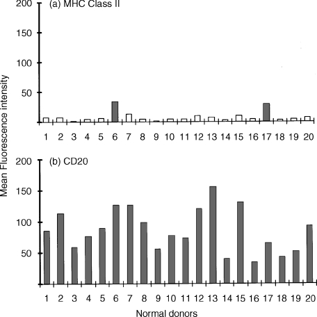
Figure 4.
Demonstration of cytoplasmic CD antigens by flow cytometry following fixation and permeabilization of neutrophils: normal donor variation. (a) Demonstration of cytoplasmic MHC Class II antigen within normal human peripheral blood neutrophils (R3) obtained from 20 normal donors. Results obtained from histogram analysis are expressed as the mean fluorescence intensity (MFI). Two donors (indicated by solid colour) were found to express significant levels (i.e. >background uptake of isotype matched mouse immunoglobulin, MFI = 6 ± 2) of cytoplasmic MHC Class II antigen. (b) Demonstration of cytoplasmic CD20 antigen within normal human peripheral blood neutrophils (R3) obtained from the same 20 normal donors measured in parallel. Results obtained from histogram analysis are expressed as the MFI. All normal donors were found to express significant (i.e. >background uptake of isotype-matched mouse immunoglobulin, MFI = 6 ± 2] but variable levels of cytoplasmic CD20. Essentially similar results were found for CD21, CD22, CD80 and CD86.

