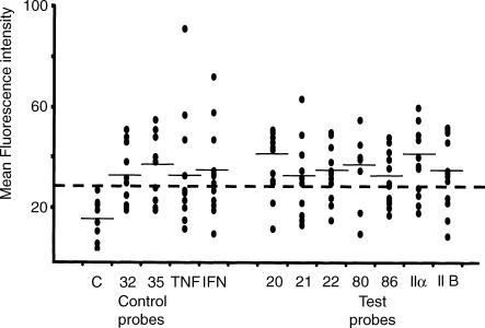
Figure 6.
In situ hybridisation flow cytometry (ISH-FC) assay: binding of control and test probes to normal human peripheral blood neutrophils. Results derived from histogram analysis, as shown in Figure 5(a), were expressed as the mean fluorescence intensity (MFI). These MFI values were plotted as a scatter diagram to show the full range of binding to neutrophils (R3) observed with 12 normal donors. Some values were identical and appear as a single dot. The average MFI value is indicated by a horizontal bar. Values greater than the upper limit for binding of the negative probe, as indicated by the dotted line, were considered significant. Control oligonucleotide probes used in all experiments were as follows: negative control probe, a sequence not recognized in human gene database (this DIG-labelled probe was used to indicate background fluorescence in this assay); probes specific for CD32 (FcγRII a-isoform) and CD35 (CR1) (cell receptor molecules known to be expressed on the surface of human neutrophils); and probes specific for TNF-α and IFN-γ (cytokines known to be synthesized by activated neutrophils).

