Figure 1.
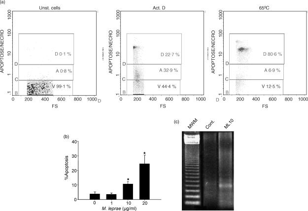
Mycobacterium leprae induces cell death in monocyte-derived macrophages of leprosy patients. (a) Flow cytometry histograms demonstrate standardization of 7-AAD staining from one representative experiment. Unstimulated PBMC (Unst. cells, left panel), PBMC stimulated with Act.D for 20 hr (middle panel), or heated at 65° for 10 min (right panel) were used to set the gates for viable (V), apoptotic (A, 7-AADdim staining), and dead cells (D, 7-AADbright), respectively. Negligible 7-AAD incorporation was seen in the live cells. (b) Evaluation of apoptosis was assessed by flow cytometry after dual staining with 7-AAD and anti-CD14 antibody in cultures stimulated or not with M. leprae 1, 10, or 20 μg/ml for 2 days. Values represent mean percentage apoptosis ± SEM of five different experiments. *Significant differences (P < 0·05) when compared to unstimulated cultures or cultures stimulated with M. leprae 1 μg/ml. (c) Agarose gel electrophoresis showing fragmented DNA in cultures stimulated or not (Cont) with M. leprae 10 μg/ml. The 123 bp DNA ladder was used as the standard molecular weight marker (MWM).

