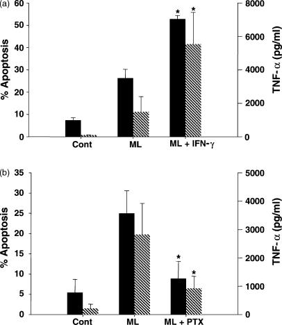
Figure 3.
Modulation of Mycobacterium leprae-induced apoptosis and TNF-α levels in monocyte-derived macrophages obtained from leprosy patients in vitro. (a) Flow cytometry analysis (%) of apoptosis (solid bars) was evaluated in the PBMC cultured for 2 days and stimulated or not (Cont) with M. leprae (ML) 20 μg/ml, in the presence or absence of IFN-γ 100 U/ml (a), after dual staining with 7-AAD and anti-CD14 antibody. The rate of cell death and TNF-α values (hatched bars), measured in these same culture supernatants by ELISA, showed (*) significant differences when IFN-γ was added to the wells (P < 0·05). Results are presented as mean ± SEM of five and three individual experiments, respectively. (b) Following stimulation of the PBMC with M. leprae in the presence or absence of PTX, apoptosis and TNF-α release were evaluated as described. *Significant differences when compared to the M. leprae-stimulated cultures. Results are mean ± SEM of four different experiments. Background values for apoptosis and TNF-α detected in the PBMC cultured with PTX alone were 7·4 ± 5·5% and 85·7 ± 25·8 pg/ml.

