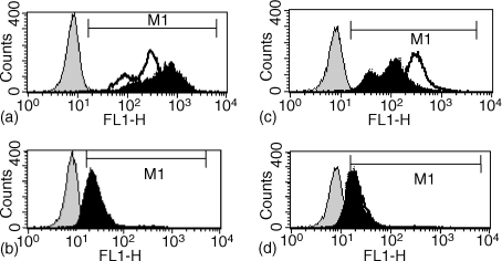
Figure 4.
Effect of ES-62 exposure in vivo on BCR-promoted ErkMAPkinase activation of spleen cells ex vivo. Splenic mononuclear cells derived from mice pre-exposed either to PBS or ES-62 (0·2 µg/hr) were stimulated with medium or F(ab′)2 fragments of anti-IgM (50 µg/ml) for 10 min before processing for flow cytometric analysis of Erk or active, dually phosphorylated Erk expression as described in Materials and Methods. In (a) and (b), PBS-exposed cells were stimulated with medium or F(ab′)2 fragments of anti-IgM and analysed for phosphoErk (a) or Erk (b) expression. In (c) and (d), ES-62-exposed cells were stimulated with F(ab′)2 fragments of anti-IgM and analysed for phosphoErk (c) or Erk expression (d). In all cases, the antibody control plot represented samples stained with rabbit IgG followed by the FITC-conjugated secondary antibody and the M1 gate was set at the 1% cut-off point of the negative cell population. MFI represents mean fluorescence intensity.

