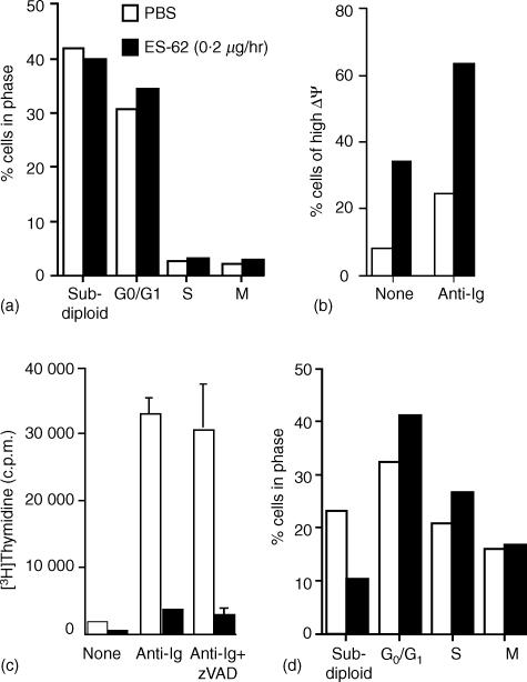
Figure 5.
Pre-exposure to ES-62 does not induce antigen receptor driven apoptosis of B lymphocytes. In (a), splenic mononuclear cells (106) from PBS or ES-62-treated mice were stimulated for 48 hr with F(ab′)2 fragments of anti-IgM before determining DNA content by propidium iodide staining and FACS analysis as described in Materials and Methods. In (b), splenic mononuclear cells (106) from PBS or ES-62-treated mice were stimulated for 24 h in the presence of media or F(ab′)2 fragments of anti-IgM (anti-Ig) before determining mitochondrial potential. Mitochondrial potential was determined by DiOC6(3) staining and FACS analysis as described in Materials and Methods. Data represents DiOC6(3) high (non-apoptotic) cell populations as determined on a logarithmic FL-1 axis and expressed as a percentage of the total number of cells analysed. Proliferation of splenic mononuclear cells from PBS or ES-62-treated mice was assessed by measurement of DNA synthesis (c). Cells (2 × 10⁵ cells/well) were treated for 48 hr with media or F(ab′)2 fragments of anti-IgM (anti-immunoglobulin) in the presence and absence of the pan-caspase inhibitor zVAD-fmk (10 µm). The levels of [3H]thymidine incorporation into DNA were measured and the data expressed as means ± SD (n = 3). In (d), splenic mononuclear cells (106) from PBS or ES-62-treated mice were stimulated for 24 h with F(ab′)2 fragments of anti-IgM before determining DNA content of the B220+ B-cell population by propidium iodide staining and FACS analysis as described in Materials and Methods. All data are representative of at least three independent experiments.

