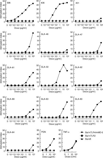
Figure 3.
NF-κB activation in Ba/F3 cells in response to various synthetic disaccharide and monosaccharide lipid A analogues. Cells were stimulated with the indicated doses of each test specimen in RPMI-1640 supplemented with 10% FBS at 37° for 4 hr. Luciferase activity in the cell lysate was measured. Results are shown as relative luciferase activity, which is a ratio of stimulated activity to non-stimulated activity, in each cell line. Similar results were obtained from three independent experiments. Peptidoglycan (PGN) and TNF-α were used as positive control specimens in this experiment.

