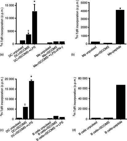
Figure 2.
DC but not Mφ or naïve B cells present ISCOMs OVA to antigen specific CD4+ T cells in vitro and this is enhanced by exposure to LPS. (a and c) BM DC, BM mφ or naïve B cells were pulsed with 10 µg/ml OVA ISCOMs for 2 hr, or pulsed with antigen and then stimulated with LPS ± IFN-γ for 24 hr before washing and culture with DO11.10 lymphocytes for 48 hr. (b) Resting BM mφ or (d) naïve B cells were pulsed with 10 µg/ml OVA ISCOMs or 10 µg/ml OVA peptide for 2 hr, washed and cultured with DO11.10 lymphocytes for 48 hr. The results shown are mean 3H-TdR incorporation ± 1 sd from triplicate cultures. (*P < 0·01 versus DC not treated with LPS, †P < 0·01 versus ISCOMs pulsed B cells or Mφ).

