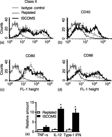
Figure 4.
Activation of DC by ISCOMs. BM DC were plated in the absence or presence of 10 µg/ml OVA ISCOMs for 4 h and the expression of (a) class II MHC (b) CD40 (c) CD80 and (d) CD86 was assessed on CD11c+ DC by flow cytometry. (e) RNA was extracted from the DC, reverse transcribed, and the relative amount of TNF alpha, IL-12 and type-1 IFN (IFN-β) determined by real time PCR and normalized to HPRT levels in each sample. Determinations were made in triplicate and mean ± 1 SD were determined. (*P < 0·01 versus controls).

