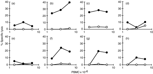
Figure 1.
Lysis of Mycobacterium bovis-infected macrophages (Mφs) by cytotoxic cells. Peripheral blood mononuclear cells (PBMC) from two calves (no. 1 and no. 2), experimentally infected with M. bovis 11 weeks previously, were restimulated in vitro and assayed for cytotoxic T-lymphocyte (CTL) activity on M. bovis-infected (•) or uninfected (○) Mφs. PBMC from calf no. 1 were cultured alone (a), with 6 × 10⁵ colony-forming units (CFU) of M. bovis (b), with 3 × 10⁶ CFU of M. bovis (c), or with 1·5 × 107 CFU of M. bovis (d) PBMC from calf no. 2 were cultured with bovine purified protein derivative (PPD) (e), with 6 × 10⁵ CFU of M. bovis (f), or with M. bovis-infected Mφs (g) or uninfected Mφs(h).

