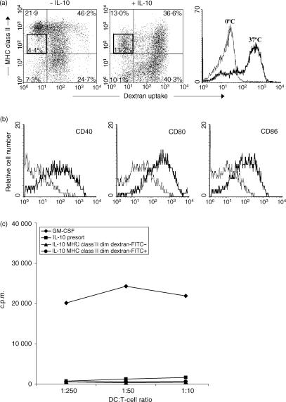
Figure 5.
Antigen-uptake capacity of dendritic cells (DC), generated by culture with granulocyte–macrophage colony-stimulating factor (GM-CSF), with or without interleukin-10 (IL-10). DC were cultured with GM-CSF, with or without IL-10. On day 9, DC were incubated with dextran–fluorescein isothiocyanate (FITC) for 1 hr at 37°, or at 0° as a control. Afterwards, the cells were washed twice with ice-cold medium. (a) Cells were stained for major histocompatibility complex (MHC) class II expression and analysed on a flow cytometer, using 7-aminoactinomycin D (7-AAD) to exclude dead cells. (b) Expression of CD40, CD80 and CD86 on gated MHC class IIdim dextran–FITCnegative cells (thin line) and on gated MHC class IIdim dextran–FITCpositive cells (thick line) was analysed by four-colour flow cytometry (dextran–FITC/CD40– CD80– or CD86–phycoerythrin (PE)/7-AAD/MHC class II biotin + streptavidin-allophycocyanin (APC)]. (c) MHC class IIdim dextran–FITCnegative cells and MHC class IIdim dextran–FITCpositive cells were sorted using a FACScalibur. Graded numbers of both sorted and the presort DC were cultured with purified allogeneic CD4+ T cells. Proliferation was assessed after 4 days by measuring the incorporation of [3H]thymidine.

