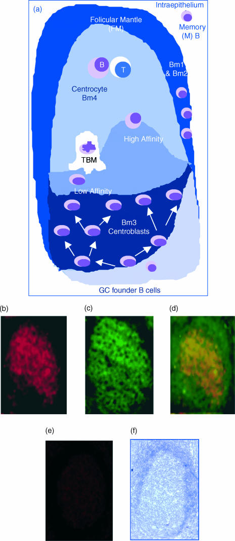
Figure 1.
Expression of interleukin (IL)-8 within germinal centres (GCs) by immunohistology. (a) A schematic representation of the GC reaction: an immunoglobulin M (IgM)-expressing GC founder migrates to the dark zone of a GC and undergoes clonal expansion and affinity maturation by somatic hypermutation. The mutated cells migrate to the basal light zone, where they are subjected to antigen-dependent selection. High-affinity (High) cells are selected and migrate to the apical light zone of GCs, where they encounter and present antigen (Ag) to T cells. Low affinity (Low) centroblasts undergo programmed cell death and are subsequently cleared by tingible body macrophages (TBM). (b) Reactivity of frozen tonsil (acetone-fixed) tissue sections to an affinity-pure anti-IL-8 immunoglobulin G (IgG). (c) B-cell identification by specific reactivity to an anti-CD20 monoclonal antibody (mAb). (d) Co-localization of the red and green fluorescence (anti-CD20), indicating the expression of IL-8 by CD20 B cells, as depicted by the yellow/orange merge. (e) Lack of reactivity to a non-immune IgG, used as a negative control. (f) Integrity of the tissue sections used, as determined by Gill's haematoxylin staining.

