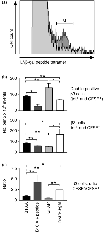
Figure 5.
Analysis of transferred, 5(6)-carboxyfluorescein diacetate N-succinimidyl ester (CFSE)-labelled β3 cells. Where indicated, the mice received 300 µg of β-galactosidase (β-gal) peptide, in saline, intraperitoneally (i.p.), 3 days post-transfer. Splenocytes were harvested on day 13 and stained with anti-CD3+, anti-CD8+, and antigen-presenting cell (APC)-labelled Ld/β-gal peptide tetramer. CD3+ CD8+ T cells were identified by fluorescence-activated cell sorter (FACS) and then analysed for Ld/β-gal peptide tetramer staining and CFSE content. (a) Plot of tetramer staining on splenocytes from mice that did (dark grey) or did not (light grey) receive β3 cells. The (M) bracket represents tetramer-positive cells, which are β3 cells that are either CFSE+ or CFSE−. (b) Number of tetramer-stained β3 cells recovered from the transgenic (Tg) and control mice. The mean result is shown of four animals and six determinations. tet+, tetramer positive. (c) Ratio of CFSE− : CFSE+β3 cells from analyses, as shown in (b). The average of all experiments is shown. Significance was determined by the t-test; comparisons are indicated by the brackets. *P < 0·05; **P < 0·01.

