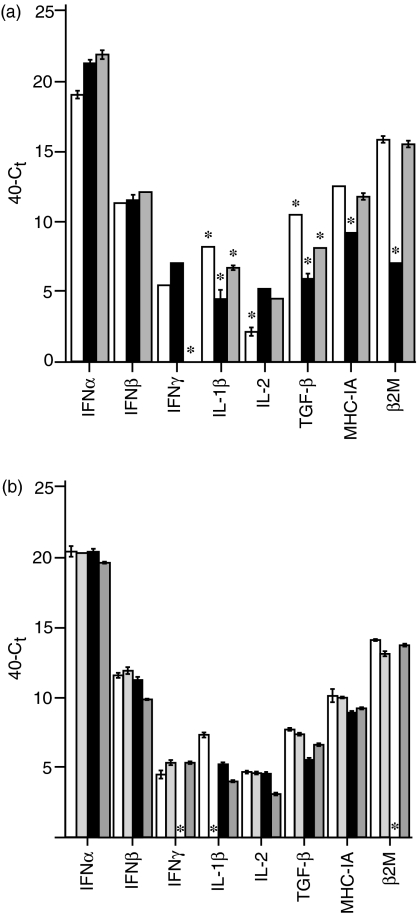
Figure 2.
Constitutive transcription of IFN-α, IFN-β, IFN-γ, IL-1β, IL-2, TGF-β4, MHC IA and β2M in unstimulated thymocytes. Transcript is quantified as 40–Ct and standardized against a 28S RNA standard. Significant differences in measured transcript are indicated by a star above the sample. (a) Transcript levels measured at E14 (unshaded), E18 (black shading) and D1 (grey shading). (b) Transcript levels in CD4+ TCR+ (unshaded), CD8+ TCR+ (light grey), CD4+ TCR− (black) and CD8+ TCR− (dark grey) subsets at E18.

