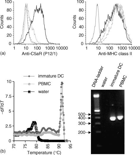
Figure 1.
Immature monocyte derived dendritic cells express the C5aR on the protein level (a) and mRNA level (b). (a) Binding of anti C5aR monoclonal antibody P12/1 to immature DC as determined by fluorescence cytometry. The histogram of the isotype control (thin line) and C5aR monoclonal antibody P12/1 binding (thick line) is shown. The specifity of binding was tested by preincubation of the antibody P12/1 with a 20fold weight excess of the peptide EX1 (dotted line), which had been used for generating the antibody, and by preincubation of the cells with C5a (dashed line). The preincubation prevented the binding of P12/1 to the C5aR. Anti-MHC class II antibody staining was not altered by preincubation of the antibody with EX1 peptide or preincubation of cells with C5a. (b). Detection of C5aR in immature DC by LightCycler RT-PCR. LightCycler melting curve analysis showed the specific peak for C5aR, which is clearly distinct from the peak caused by primer-dimer formation visible in the water (= negative) control. PBMC were used as positive control. Analysis of LightCycler PCR products by agarose gel electrophoresis revealed bands of the expected size (381 bp).

