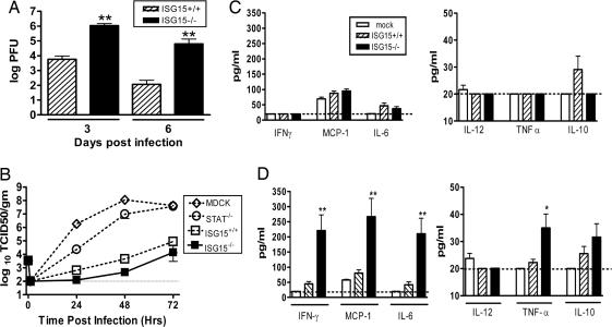
Fig. 2.
Characterization of influenza pathogenesis in ISG15−/− mice. (A) Lung homogenates from ISG15+/+ (hatched bars) or ISG15−/− (filled bars) mice infected with influenza B/Lee virus at 1 × 10⁶ pfu i.n were titered by plaque assay. Error bars represent SEM. Fifteen to 17 animals were harvested for each time point. (B) Growth of influenza A rWSN was determined on Madin–Darby canine kidney (MDCK) cells (open diamonds), STAT1−/− (open circles), ISG15+/+ (open squares), or ISG15−/− (filled squares) murine embryonic fibroblasts (MEFs). Data are representative of two independent experiments. Error bars represent SEM. (C and D) Serum cytokine levels were analyzed at 3 (C) or 6 (D) days postinfection in mock-infected (open bars), ISG15+/+ (hatched bars), or ISG15−/− (filled bars) mice infected with 1 × 10⁶ pfu influenza B/Lee virus i.n. ∗, P < 0.001; ∗∗, P < 0.0001.

