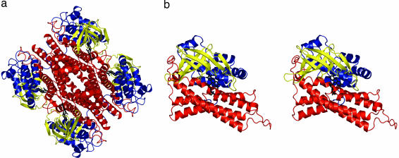
Fig. 2.
Overall structure of C2. (a) Ribbon diagram of the 222 symmetric C2 tetramer. N-terminal domains (residues 24–143) are in blue, β-sheet domains (144–237) are in yellow, and C-terminal domains (238–422) are in red. FMNH− is in black sticks. (b) Ribbon stereo diagram of the C2 monomer. This view is obtained from rotating the top monomer of a by 45° around the vertical axis. Produced with PyMOL [DeLano, W. L. (2002) www.pymol.org].

