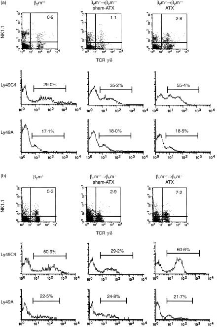
Figure 2.
Expression of Ly49 receptors on NK1.1+ γ/δ T cells in the liver of BM-transplanted chimera mice. BM chimera mice were generated by transplanting T-cell-depleted BM from β2m−/− mice into lethally irradiated 8-week-old B6-Ly5.1 mice [β2m−/−→β2m+/+ (a)] or BM from B6-Ly5.1 into β2m−/− mice [β2m+/+→β2m−/− (b)] either under euthymic (sham-ATX) or athymic (ATX) conditions. Sixteen to 20 weeks after BM transplantation, the proportions of host-derived cells were checked by the negative expression of Ly5.1 for β2m−/−→β2m+/+ chimeras and by the positive expression for β2m+/+→β2m−/− chimeras. For four-colour FACS analysis, liver MNCs from naive β2m+/+ or β2m−/− mice were stained with purified-anti Ly5.1 mAb (A20) followed by Cy5-anti-mouse IgG. After vigorous washing, cells were stained with PE-anti-NK1.1 mAb (PK136), biotin-anti-γ/δ TCR mAb (GL3) and FITC-anti-Ly49C/I mAb (5E6), or FITC-anti Ly49A mAb (A1) followed by streptavidin-RED613™. Chimera mice in which the percentage of donor-derived cells was proven to be 95% or more were used for analysis. Expressions of NK1.1 and γ/δ TCR are displayed by two-dimensional dot plots, and the expressions of Ly49C/I and Ly49A on NK1.1+ and γ/δ TCR+ cells are shown as single histograms. Representative data from three to five mice in each group are shown as typical profiles.

