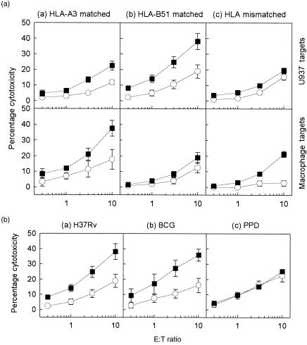
Figure 3.
(A) Mycobacterium tuberculosis-specific cytolysis generated by the (a) human leucocyte antigen (HLA)-A3-matched (n = 4; TS, PB, ES and EC), (b) HLA-B51-matched (n = 2; RG and BR), and (c) HLA-mismatched (n = 6; MH, NP, SG, SJ, MF and WM) donor cytotoxic T lymphocytes (CTL) against U937 (top panel) and autologous macrophage (Mφ) targets (bottom panel). (B) Mycobacterial antigen-specific cytolysis generated by HLA-B51-matched donor CTLs (n = 1; RG) against U937 targets infected with (a) M. tuberculosis H37Rv (5 colony-forming units [CFU]/cell) (b) bacillus Calmette–Guérin (BCG) (5 CFU/cell), or (c) pulsed with the soluble mycobacterial extract purified-protein derivative (PPD) (3 µg/ml). Target cells were infected with M. tuberculosis (5 CFU/cell) (▪) or pulsed with an irrelevant antigen, streptokinase-streptodornase (SK-SD) (○). Cytotoxicity against U937 and autologous Mφ targets was measured after 4 hr. Each data point represents the mean percentage cytolysis (± SEM) of at least three independent experiments.

