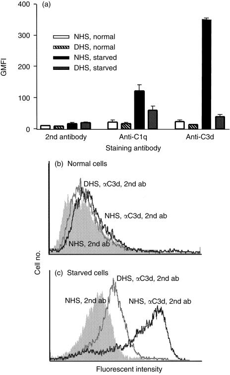
Figure 3.
Complement activation by apoptotic HUVEC. HUVEC were cultured overnight in complete EGM (normal) or EGM without serum or growth factors (starved). Cells were incubated with 10% NHS or DHS for 30 min at 37°, washed and stained with mAb specific for C1q or C3d and PE-conjugated goat anti-mouse IgG (second antibody). (a) The GMFI values for cells treated with NHS or DHS and of cells stained with second antibody only are shown. The results shown are the means ± SEM of two experiments. (b,c) Representative histograms of normal and starved cells incubated with NHS (dark line) or DHS (grey line) and stained with anti-C3d and second antibody compared to cells incubated with NHS and stained with second antibody alone (shaded histogram).

