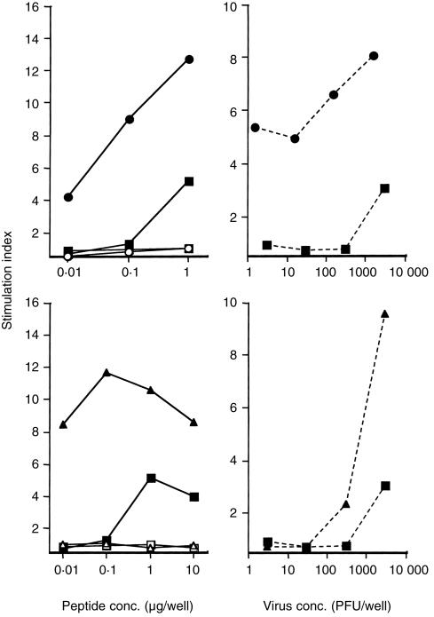
Figure 5.
Immunogenicity of synthetic peptide HA: 307–319 after immunization onto bare skin with LT (top panel) and CT (bottom panel). Splenocytes collected after two administrations of peptide alone (square) or with the mucosal adjuvants LT (circle) or CT (triangle) were cultured in the presence of increasing concentrations of homologous peptide (▪, •, ▴ for mice immunized with peptide, peptide + LT, peptide + CT, respectively), irrelevant peptide (□, ○, ▵ for mice immunized with peptide, peptide + LT, peptide + CT, respectively; left top and bottom panel) or heat-inactivated influenza virus (▪, •, ▴ for mice immunized with peptide, peptide + LT, peptide + CT, respectively; right top and bottom panel). Supernatants collected after 72 hr of culture were tested for their ability to secrete IL-2 measured using the CTLL-2 cells. The data represent the mean SI values from triplicate cultures. Background values in the absence of antigen were 645 c.p.m. (for peptide and virus) after immunization with peptide alone, 774 c.p.m. and 905 c.p.m. (for peptide and virus, respectively) after immunization with peptide and LT, and 890 c.p.m. (for peptide and virus) after immunization with peptide and CT. Standard deviations in triplicate cultures were < 20%.

