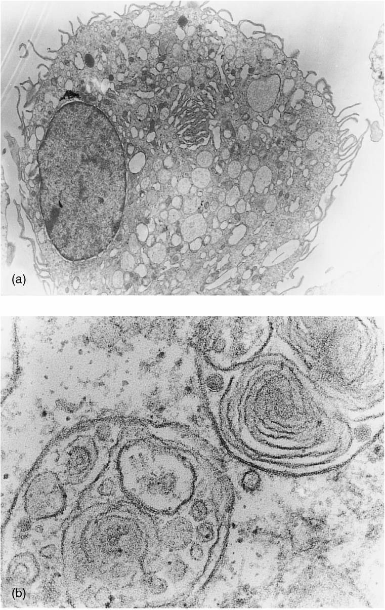
Figure 1.

Transmission electron microscopy was used to evaluate the cellular morphological characteristics of adherent peripheral blood mononuclear cells (PBMC) after 7 days of culture in the presence of porcine granulocyte–macrophage colony-stimulating factor (GM-CSF) and interleukin-4 (IL-4). (a) Electron micrograph showing a cell with a large diameter and microvillous projection of the plasma membrane (magnification ×2600). (b) Details of the multivacuolar and multilamellar intracytoplasmic organites (magnification ×46000).
