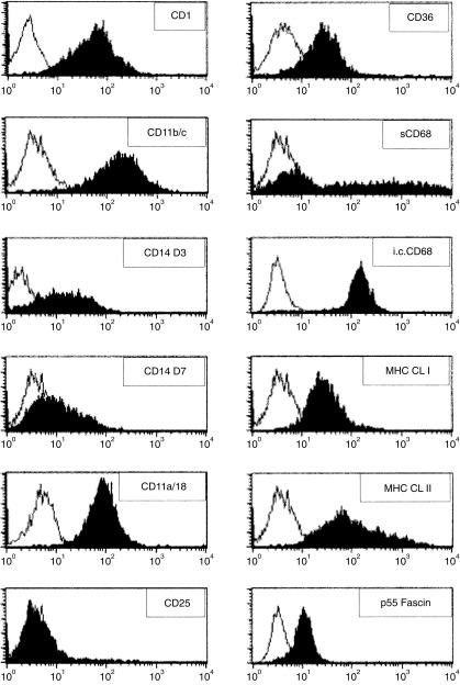
Figure 2.
Flow cytometric phenotyping of the porcine dendritic-type cell. Cells were stained with control isotypes (open histogram) or with the indicated anti-human or anti-porcine monoclonal antibody (mAb) against cell surface markers (solid histogram). Intracellular (i.c.) staining was performed for the human (hu) p55 Fascin. Both i.c. and surface (s) staining were performed for CD68. Data are representative of at least three experiments performed.

