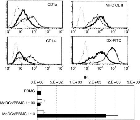
Figure 5.
Phenotypic and functional modification induced by treatment of the monocyte-derived dendritic cells (MoDCs) with IRP necrotic factors. (a) Five-day-old MoDCs were cultured for 4 days with (thick line) or without (thin line) IRP necrotic factor (1:2 dilution). Untreated (thin line) and necrotic factor-treated (thick line) MoDCs were stained for CD1a, CD14 and major histocompatibility complex (MHC) class II molecules and control isotypes (dotted line). Untreated (thin line) and necrotic factor-treated (thick line) MoDCs were incubated for 4 hr with dextran-fluorescein isothiocyanate (DX-FITC) at 39° or on ice (dotted line). (b) Proliferative responses of 2 × 10⁵ porcine PBMC in response to allogeneic untreated (open histogram) or necrotic factor-treated (solid histogram) MoDCs. IP, index of proliferaion (see Materials and methods).

