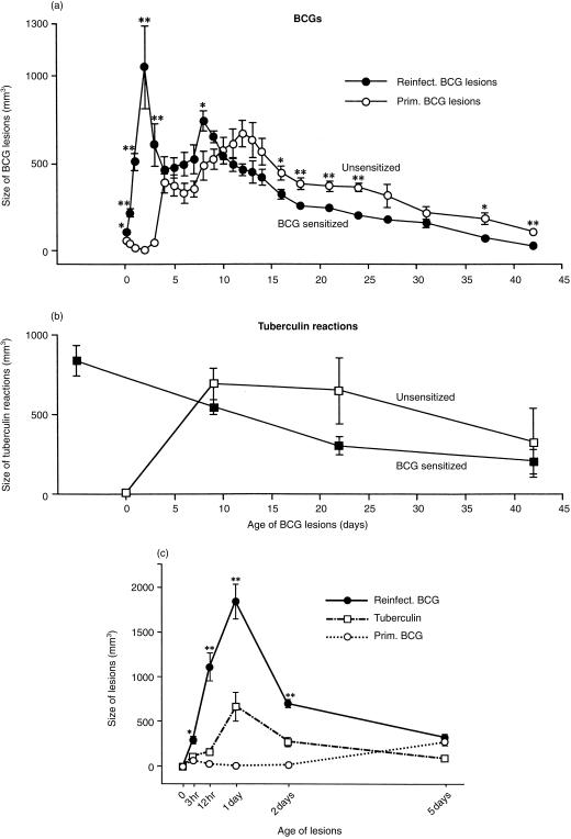
Figure 1.
(a) Size of primary BCG lesions and reinfection BCG lesions from 3 hr to 42 days in rabbits of Expt I. The reinfected rabbits had been sensitized intradermally by BCG 24 days previously. The reinfection BCG lesions were many times larger than the primary BCG lesions at 3, 12, 24 and 48 hr, a fact that was apparently initiated by an antigen–antibody reaction (see text). Also, the size of the reinfection BCG lesions reached a second peak at 8 days, whereas the primary lesions reached a similar peak at 12 days. These second peaks were apparently caused by an antigen-specific CMI/DTH reaction (see text). After the second peaks, the lesions slowly regressed. Each point represents the mean of lesions from five rabbits and its standard error. (b) Size of 2-day tuberculin reactions in rabbits of Expt I. In the reinfected host, tuberculin sensitivity was highest before challenge. This sensitivity declined thereafter, and no booster effect from the second BCG injection was apparent. In contrast, in hosts with primary BCG infections, tuberculin sensitivity was strong by 9 days and tended to remain higher than that present in the reinfected hosts, possibly because the infecting bacilli were not destroyed as readily. Each point represents the mean of five rabbits and its standard error. (c) Size of primary and reinfection BCG lesions and size of tuberculin reactions 3 hr to 5 days old in Expt II, from which tissue sections were obtained and evaluated. As in Expt I, the reinfected rabbits were sensitized intradermally by BCG 24 days previously. During these first 5 days, the size of the reinfection BCG lesions and of the tuberculin reactions followed the same pattern. Each point represents the mean of at least four lesions with its standard error. In (a) and (c), reinfection BCG lesions versus primary BCG lesions: *P < 0·05 and **P < 0·01.

