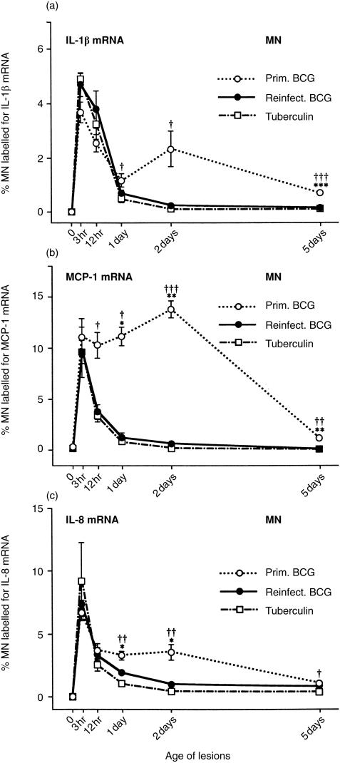
Figure 6.
Percentage of MN labelled for (a) IL-1β mRNA, (b) MCP-1 mRNA and (c) IL-8 mRNA in the three types of lesion. The percentage of MN containing the three cytokine mRNAs shows early peak levels at 3 hr. Then, this percentage of MN rapidly declines in BCG lesions of reinfection and in tuberculin reactions, but remains relatively high in primary BCG lesions at 2 days, especially the percentage containing MCP-1 mRNA. At 2 days, the primary lesions are growing in size, whereas the reinfection lesions and tuberculin reactions are regressing (see Figure 1c). Each point represents the mean of four lesions with its standard error: for reinfection BCG lesions versus primary BCG: *P < 0·05, **P < 0·01 and ***P < 0·001; for tuberculin reactions versus primary BCG lesions: †P < 0·05, ††P < 0·01 and †††P < 0·001.

