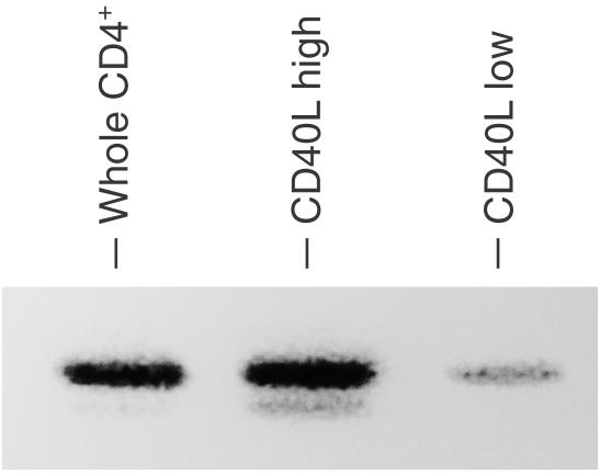
Figure 6.
Syk expression on patient's CD4+ T cells that express high levels of CD40L. Purified CD4+ T cells of the patient were stimulated with biotinylated anti-CD3 plus streptavidin for 3 hr. The cells were stained with PE-conjugated anti-CD40L antibody and were sorted into CD40L+ (CD40L high) and CD40L– (CD40L low) populations using FACS Vantage. Cell lysates of the sorted cells (2 × 10⁶ each) were analysed by SDS–PAGE and were subjected to anti-Syk blots.

