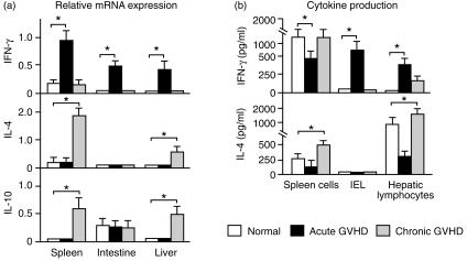
Figure 7.
Th1 and Th2 cytokine expression in the target organs. (a) Total RNA was isolated from tissue samples and RT-PCR cDNA products were generated and amplified using specific primers for IFN-γ, IL-4, IL-10 and β-actin. The amounts of the IFN-γ, IL-4 and IL-10 RT-PCR cDNA products were normalized by the β-actin control. Data represent the mean±SD of the relative expression of RT-PCR cDNA products in four mice; *P < 0·05. (b) Spleen cells, intestinal IEL and hepatic lymphocytes were prepared as described in the Materials and Methods. The cells (1 × 10⁵/well/200 µl) were incubated for 72 hr on anti-CD3 mAb-coated plates that had been made by preincubation of each plate with 20 µg/ml of 145-C11 in PBS overnight at 4°. The concentrations of IFN-γ and IL-4 in the culture supernatants were measured by ELISA using anti-mouse-IFN-γ and IL-4 mAbs, respectively. Data represent the mean±SD for four mice. *P < 0·05.

