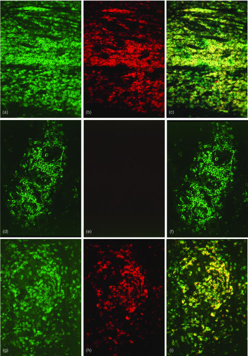
Figure 3.
Double immunofluorescence analysis of CD3ε and CD3ζ expression in situ. Hyperplastic tonsil (a, b, c), a tuberculoid leprosy skin biopsy (d, e, f) and a sarcoid lymphnode (g, h, i) were stained for CD3ε (green fluorescence, a, d, g), CD3ζ (red fluorescence, b, e, h) and double exposed for colocalization of both CD3ε and CD3ζ (yellow fluorescence, c, f, i). In hyperplastic tonsil, CD3ζ expression colocalized 100% with CD3ε expression. In the tissue area chosen close to endothelium in this example, several cells exhibited CD3ε positivity and CD3ζ negativity. In an example of tuberculoid leprosy, CD3ε positive cells were detected throughout the granuloma (d) whereas CD3ζ expression was barely visible (e), resulting in no detection of CD3ε CD3ζ double positive cells (no yellow fluorescence (f). The sarcoid lymph node lesion exhibited CD3ε (g) and CD3ζ (h) expression to approximately the same extent. All CD3ζ cells were CD3ε positive (yellow fluorescence) and only a minor number of cells were CD3ε positive CD3ζ negative (i, green fluorescence).

