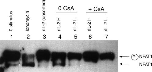
Figure 6.
Recombinant IL-2 induces CsA-susceptible NFAT1 dephosphorylation in CD154/CD40L-expressing T lymphoblasts. T lymphoblasts were cultured in the presence of rIL-2 (lanes 3–7) or left unstimulated (lane 1). CsA (5 µg/ml) was added to a subset of rIL-2-stimulated cells (lanes 6,7). After a 24-hr culture, surface expression of CD154/CD40L by stimulated lymphoblasts was assessed using indirect immunofluorescence and FACS analysis. Lymphoblasts were sorted into rIL-2-stimulated populations expressing high levels of CD154/CD40L (rIL-2H, designating 20% of the cells with the highest expression) (lanes 4,6), and populations expressing no detectable CD154/CD40L (rIL-2L) (lanes 5,7). Cells were then lysed, and the NFAT1 activation state was assessed by Western blotting. The positions of the inactivated (phosphorylated) and activated (non-phosphorylated) forms of NFAT1 are indicated to the right of the blot. Lysates from lymphoblasts which had been stimulated with ionomycin (and showed substantial NFAT1 dephosphorylation) were used as a positive control (lane 2). Shown are representative results from one of three experiments.

