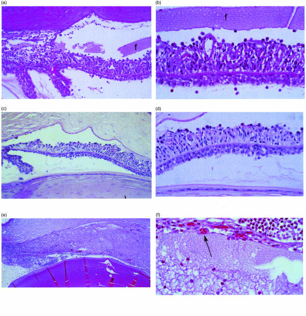
Figure 3.
(a) Anterior chamber of rat injected into the aqueous humor with 5I2 F(ab′)2 fragments. Marked inflammation of the iris and anterior chamber, equivalent to that of intact 5I2 IgG, is seen. Moderate fibrin (f) is seen in the anterior chamber (original magnification ×200). (b) High-power view of (a) showing thickening and inflammatory cells throughout the iris. The overlying fibrin is seen across the entirety of the field (original magnification ×400). (c) The anterior chamber injected identically with anti-OX-18 F(ab′)2 fragments. Inflammation is minimal and proteinaceous material is absent (original magnification ×200). (d) High-power view of (c) showing few inflammatory cells on the anterior iris surface (original magnification ×400). (e) Anterior chamber injected into the aqueous humor simultaneously with 5I2 and anti-rat CD59 monoclonal antibodies (mAbs). The inflammation totally obscures the underlying anatomy (original magnification ×100). (f) High-power view of (e) showing necrosis and thinning of iris tissue resulting from the 5I2 and anti-CD59-induced inflammation (original magnification ×400).

