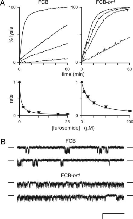
Fig. 3.
Markedly reduced sensitivity to furosemide of PSAC expressed on FCB-br1. (A) (Upper) Osmotic lysis kinetics in proline at 20°C in the presence of 0, 10, 25, and 200 μM furosemide (top to bottom traces in each graph, respectively). Proline uptake by FCB-br1 is less well inhibited by furosemide. (Lower) The corresponding dose responses for inhibition. Symbols represent the mean ± SEM lysis rates normalized to 1.0 without furosemide, calculated as described in ref. 23 from up to five measurements at each concentration. Solid lines represent a least-squares best fit to y = K0.5/(K0.5 + x) with K0.5 estimates of 0.8 and 32 μM for FCB and FCB-br1, respectively. (B) Single PSAC recordings in solution A with 10 μM furosemide in bath and pipette. Vm = −100 mV. (Scale bars: 100 ms and 2 pA.) Closed channel currents are indicated by dashes to the sides of each trace. Furosemide significantly reduces channel openings (downward deflections from the closed channel level) for PSAC on the FCB isolate but has a negligible effect on FCB-br1 channels when compared with their activities without inhibitor (Fig. 2).

