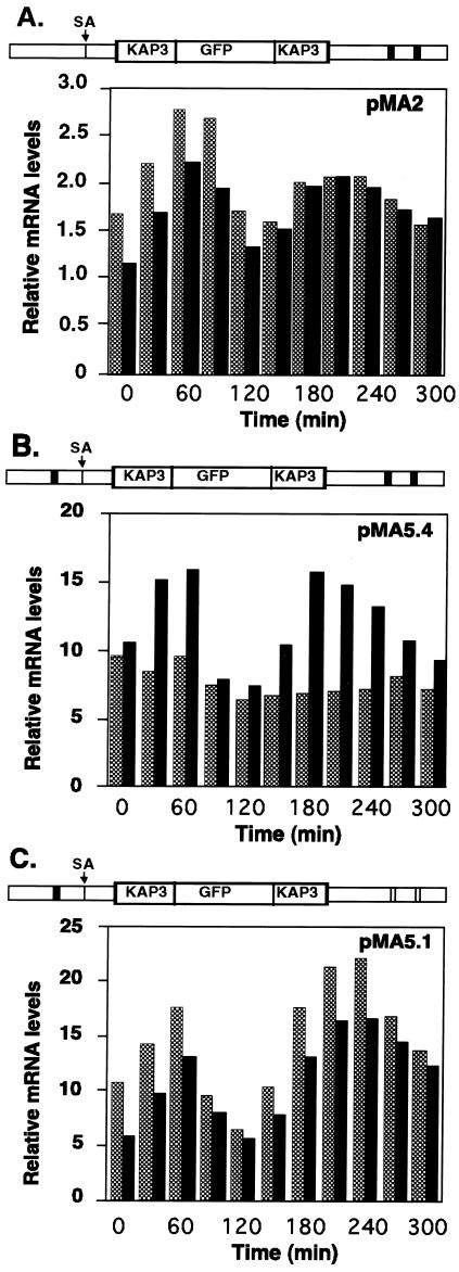
FIG. 4.
Sequence elements required for cycling of KAP3-GFP mRNA levels. Shown is a PhosphorImager quantitation of Northern blots of total RNA isolated from synchronized C. fasciculata cells carrying either pMA2 (A), pMA5.4 (B), or pMA5.1 (C). RNA samples were isolated at 30-min intervals after release from hydroxyurea arrest and analyzed as for Fig. 3. A schematic diagram of the relevant region of each plasmid, with open rectangles representing wild-type consensus octamer sequence elements (CATAGAAA) and filled rectangles representing mutated octamer sequences, is shown above the respective bar graph. Solid bars, KAP3 mRNA; dark shaded bars, KAP3-GFP mRNA.

