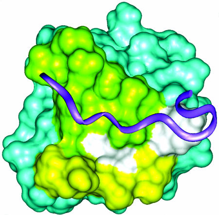
Fig. 9.
Surface view of the SH3 domain of AMIC. The region perturbed by interactions with residues in the basic region is shown in yellow, the region that is expected to be the binding site for proline-rich ligands is in green, and the overlap between these two regions is in white. The position of a hypothetical ligand (purple ribbon) is modeled by superposition of the Fyn proto-oncogen tyrosine kinase SH3 domain/ligand complex (Protein Data Bank ID Code 1A0N).

