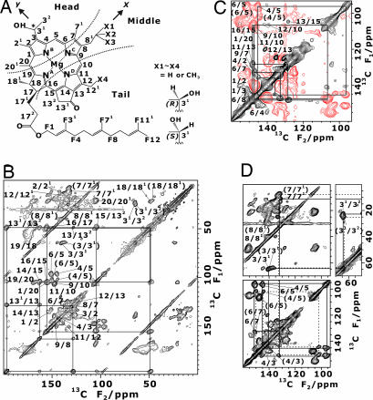
Fig. 1.
Two-dimensional MAS 13C 13C dipolar correlation NMR spectra of uniformly 13C-labeled chlorosomes from C. limicola are shown. (A) Chemical structure of BChl c isomers is shown. Numbers with prefix F are carbon atoms in the farnesyl chain. BChl c has two stereoisomers with chirality at the 31 position and homologues with different side chains at C8 and C12 as shown on the right. The head, middle, and tail parts are also indicated. The z-axis is perpendicular to the ring plane. (B) Two-dimensional 13C correlation spectrum with an rf-driven recoupling mixing time of 1.28 ms is shown. (C) Two-dimensional 13C correlation spectrum with a SPC-5 mixing time of 1.1 ms is shown. The red lines stand for negative contour levels. (D) Displayed are the connectivities of the doublet signals shown in a part of the 2D 13C NMR spectrum in B. Two individual connectivities for signals a and b are shown by solid and dashed lines, respectively, from C71 to C31, and are indicated by assignments with and without parentheses.

