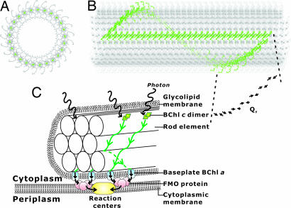
Fig. 5.
A rod element structure built with the parallel dimer layers shown in Fig. 4 E–H. (A) A top view. Magnesium atoms are colored dark green. Columns perpendicular to the page are arranged on the circumference. (B) A side view. The cylindrical structure consists of ≈1,500 molecules. A single spiral layer and a single column are colored green. A layer consisting of 25 dimers makes one spiral rotation. Twenty-nine full stacking dimers (Fig. 4 F and G) constitute the green column along the cylinder axis. The arrows represent the Qy transition dipole moments of BChls. The Qy transition dipolar vectors form an angle of 46° with the cylinder axis and an angle of 236° with the radius vector connecting Mg and the cylinder axis. (C) Schematic representation of the excitation energy transfer in a chlorosome. The green lines with arrows indicate the paths of the excitation transfer along the spiral layers to the baseplate shown in B. The model structure was taken from ref. 46.

