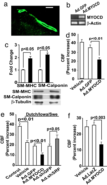
Fig. 4.
MYOCD and sh.SRF gene transfer to mouse pial arteries alters CBF responses to vibrissal stimulation. (a) Expression of GFP in cerebral pial vessels after subarachnoid infusion of Ad.GFP. (Scale bar, 50 μm.) (b) PCR for MYOCD mRNA in cerebral pial vessels after in vivo transduction with Ad.MYOCD. (c) Increase in contractile protein content in cerebral pial arteries after MYOCD gene transfer compared with GFP controls by Western blot analysis. (d–f) Effect of MYOCD gene transfer on CBF increase after whisker stimulation compared with GFP controls in wild-type (d), Dutch/Iowa/Swe (e), and APPsw+/− mice (f). In d–f, mock colony-stimulating-factor controls (vehicle) were also studied. Mean ± SEM from five animals per group.

