
Figure 4.
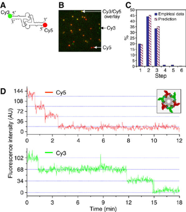
Dual-view imaging of procapsids containing both Cy3-pRNA I and Cy5-pRNA II. (A) pRNA dimer constructed with Cy3-pRNA I and Cy5-pRNA II. (B) Typical fluorescence image of procapsids bound with dual-labeled pRNA dimers. The image is an overlay from the fluorescence signals in both Cy3 and Cy5 channels, presented in pseudo-colors. Signals in the Cy3 channel were assigned green and those in the Cy5 channel were assigned red. Yellow color resulted from the overlay of red and green. Green color spots are procapsid/pRNA complexes containing Cy3 only; red color spots are the complexes containing Cy5 only. The yellow color spots are the combination of green and red, representing coexistence of Cy3-pRNA I and Cy5-pRNA II on one procapsid. (C) Comparison of empirical distribution with theoretical prediction in photobleaching steps of Cy3-pRNA I in procapsids bound with dual-labeled dimers. Prediction was based on a 70% labeling efficiency of Cy3-pRNA. (D) Fluorescence intensity versus time to show photobleaching steps of procapsids reconstituted with the dimer, elucidated in the inset showing procapsids with a pRNA hexamer composed of three Cy3-pRNA I and three Cy5-pRNA II.