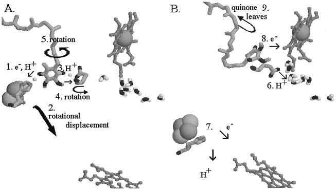
Figure 4.
Mechanism proposed for reactions at the Qo site after formation of the reaction complex. (A) The reaction complex between QH2 bound in the distal domain and ISPox bound at the interface on cyt b. The small white spheres show the H atoms of the H bonds, in both of which the quinol is the donor. The protein structure is that for the stigmatellin complex. (B) The reaction complex has dissociated to products, and the ISPred has moved to a position close to cyt c1. The protein structure is that for the myxothiazol complex. Numbers indicate the sequence of reactions. In the text, italic numbers refer to the numbered processes.

