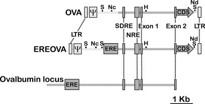
Fig. 1.
Schematic representation of the structure of the vectors described after chromosomal integration. Relative positions of lentiviral elements (light gray) and ovalbumin regulatory sequences (dark gray) are indicated, including the EIAV LTRs and packaging signal (Ψ) and the ovalbumin gene ERE; steroid-dependent regulatory element (SDRE), negative regulatory element (NRE), exon 1, intron 1, and the 5′ segment of exon 2 of the ovalbumin transcribed sequence; and the Kozak sequence and coding sequence of either miR24 or hIFNβ1a (CDS). The positions of restriction sites SpeI (S), NcoI (Nc), HindIII (H), and NdeI (Nd) used in restriction analysis are marked. The U3 region of the 3′ LTR is modified such that the integrated provirus lacks an active promoter within the LTR. Relative positions of regulatory elements in the chicken ovalbumin locus are shown.

