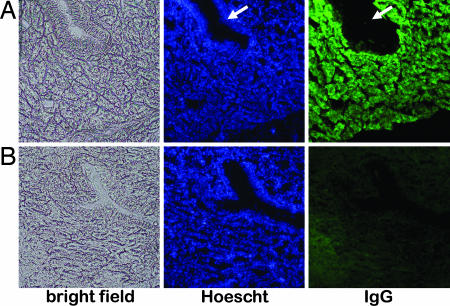
Fig. 4.
Immunohistochemical detection of miR24 in oviduct sections. (A and B) Sections of the magnum portion of the oviduct of hen OR2-4:25:59 (A) and a nontransgenic control (B) were stained with the nuclear stain Hoescht to visualize individual cells or FITC-anti-human IgG to visualize location of miR24. Tubular gland cells expressing the transgene appear green, whereas epithelial cells lining the tubular glands (indicated by arrows) do not produce miR24.

