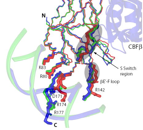
Figure 1. Comparison of structures of the apo Runt domain (1LJM, red), the Runt domain-DNA binary complex (1HJC, green), and the Runt domain-CBFβ-DNA ternary complex (1H9D, blue).

The S-switch region, βE’-F loop, and sidechains of the Runt domain residues involved in interactions with DNA are shown.
