Figure 5.
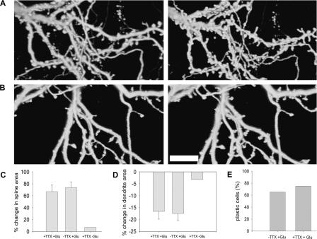
Sustained network activity is not required for glutamate-induced plasticity. (A) Three-dimensional reconstructed images of spines and dendrites in the presence of 0.5 μM TTX, before (left) and 45 min after (right) glutamate application. (B) Three-dimensional reconstructed images of spines and dendrites in the presence of 0.5 μM TTX alone before (left) and 45 min later (right). Scale bar, 10 μm. (C) The average increase in spine area following glutamate application in the presence of TTX is similar to control (67.36%; two coverslips, seven cells, 100 spines and 74.42%; six coverslips, 13 cells, 167 spines, respectively), but higher than the change in cells which were exposed only to TTX and not to glutamate (7.82%; one coverslip, three cells, 35 spines). (D) The average reduction in dendrite area following glutamate application in the presence of TTX is similar to control (16.78%; two coverslips, five cells, 25 segments and 17.8%; three coverslips, 10 cells, 30 segments respectively), but higher than the change in cells which were exposed only to TTX and not to glutamate (3.34%; one coverslip, three cells, 17 segments). (E) The percentage of plastic cells in the presence of TTX is similar to glutamate. χ2(1) = 0.09.

