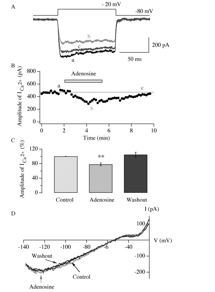
Fig.8.
Adenosine inhibits voltage-dependent calcium currents in hypocretin/orexin neurons. A, sample traces of whole-cell voltage-dependent calcium currents recorded at various stages in our experiments are shown. Voltage-dependent calcium currents were induced in hypocretin/orexin neurons held at −80 mV under voltage clamp with a voltage step from −80 mV to −20 mV. B, the time course of a typical experiment is shown. The application of adenosine (30 μM) is indicated as the filled bar above the time course curve. Letters a, b and c indicate time points when sample traces were recorded. C, pooled data from all tested neurons were analyzed and plotted, which demonstrate that the amplitude of voltage-dependent calcium currents significantly decreases in the presence of adenosine (30 μM) (**, P<0.01, ANOVA). D, the I-V relationship of membrane currents induced by a ramp pulse (from −140 mV to −20 mV, duration=600 ms) before, during and after the application of adenosine is shown. There is no significant GIRK current in the presence of adenosine.

