Figure 1.
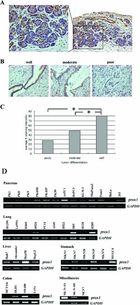
Expression of Prox1 is downregulated in human pancreatic cancer and correlates with its differentiation. (A) Immunostaining of normal pancreatic tissue. Prox1 expression is localized in ductal cells. (B) Immunostaining of pancreatic cancers of various grades: well-differentiated (well), moderately differentiated (moderate), and poorly differentiated (poor). (C) Staining intensity for Prox1 was estimated for each cell type. Columns show the relationship between tumor differentiation level and the average percent staining intensity for prox1. *P < .05. (D) Expression and mRNA editing of prox1 in diverse types of human cancer cell lines. RT-PCR analyses of prox1 expression in human pancreatic cancer cell lines: HeLa and 293 cells (pancreas), human lung cancer cell line (lung), human HCC (liver), human gastric cancer cell lines (stomach), human colon cancer cell lines (colon), and other types of human cancer cell lines (miscellaneous). GAPDH was used as internal control.

