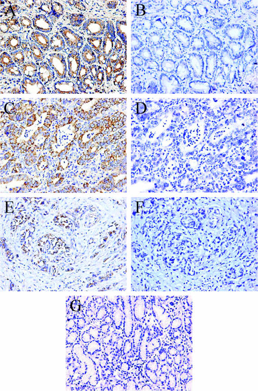
Figure 1.
Immunohistochemical staining of URG4 in normal gastric tissues and gastric cancer with different stages of differentiation. (A) Anti-URG4 staining of well-differentiated gastric carcinoma tissue. (B) Preimmune rabbit serum used to stain a consecutive section from the same patient as in (A). (C) Anti-URG4 staining of moderately differentiated gastric carcinoma tissue. (D) Preimmune rabbit serum used to stain a consecutive section from the same patient as in (C). (E) Anti-URG4 staining of poorly differentiated gastric carcinoma tissue. (F) Preimmune rabbit serum used to stain a consecutive section from the same patient as in (E). (G) Normal epithelium exhibited negative URG4 immunostaining. (A–G) Original magnification, x200.

