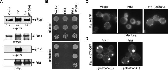
Figure 1.
Localization of phosphorylated Pan1p in vivo. (A) In vivo phosphorylation of Pan1p by Prk1p. Pan1p were precipitated with TAP-tag from cells overexpressing vector alone, Prk1 or Prk1(D158A) and immunoblotted with anti-pThr or anti-Pan1p antibody. Relative phosphorylation levels are indicated under the top panel. Prk1 and Prk1(D158A) were immunoprecipitated with anti-Myc antibody and immunoblotted with anti-pThr or anti-Myc antibody. (B) Viability of cells overexpressing Prk1 or Prk1(D158A). Cells were grown in YPD media overnight, and serial dilutions were spotted on glucose and glactose plates. (C) Localization of Pan1-GFP in cells overexpressing Prk1 or Prk1(D158A). The same cells as in B were grown to an OD600 of ∼0.3 in selective synthetic medium with 2% raffinose, and then Prk1 or Prk1(D158A) was induced with 2% galactose for 120 min. (D) Localization of Pan1-15TA-GFP in cells overexpressing Prk1. Pan1-15TA cells were grown to an OD600 of ∼0.3 in selective synthetic medium with 2% raffinose (left), and then Prk1 was induced with 2% galactose for 120 min (right).

