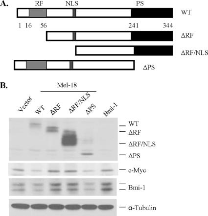
Figure 4.
Structural analysis of Mel-18. (A) Schematic representation of mutants of Mel-18 depicting various domains. These mutants were generated by PCR and cloned in the pLPC retroviral vector. (B) Stable overexpression of wild type (WT) and the mutants of Mel-18 in MCF10A cells; WT and the PS mutant down-regulated Bmi-1 and c-Myc expression, whereas overexpression of ΔRF and ΔRFNLS mutants led to up-regulation of Bmi-1 and c-Myc. WT or mutants of Mel-18 were stably expressed using retroviral expression, and Bmi-1, c-Myc, Mel-18, and α-tubulin were detected by Western blot analysis as described in Materials and Methods.

